Changelog
Timeline Summary Mode for Video Annotation
V7 Darwin now includes Timeline Summary, a new feature designed to give you a high-level overview of your video annotations. With the timeline mode enabled, you can quickly review annotation coverage, navigate large files, and streamline your review process.
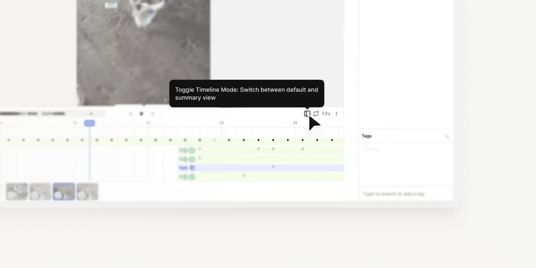
Access with Shift + D: Open Timeline Summary Mode using the new button on the right-hand side of the interface or the hotkey Shift + D.
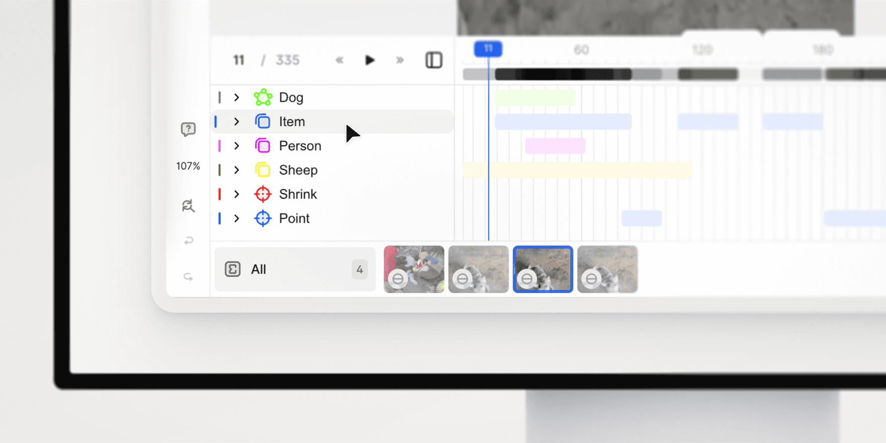
Summary by Class: Annotations are grouped by class on the left panel. Expand any class to view individual annotations and their frame ranges.
Click to Jump: Click once on an annotation to navigate to it, or double-click to open it in the regular timeline.
Read-Only Mode: The summary timeline is read-only to prevent accidental edits. However, you can continue to edit annotations in the canvas and layer bar.
Clean View: Keyframes and detailed elements are hidden from the summary view, providing a clear, scannable overview.
Heatmap Overview: A heatmap at the top of the timeline shows annotation density across the video, with the zoomed-in section highlighted in blue for easy orientation.
Timeline Summary Mode makes it easier to navigate, audit, and review video annotations, especially for large or complex files.
Learn more: V7 Darwin Documentation

Apr 25, 2025
Flood Fill Tool Now Available in V7 Darwin
This update lets you annotate volumetric regions in CT/MRI scans based on intensity similarity of connected voxels or pixels.
You can adjust tolerance levels and create annotation masks in just a few clicks. It works similarly to the flood filling functionality of Slicer 3D or to magic fill/paint bucket tools in image editing software like Photoshop. If you are familiar with these tools, you’ll feel right at home.
Key functionalities:
Switch between 3D or 2D fill for voxel or pixel-based masks
Adjust tolerance level to label similar regions automatically
Combine Flood Fill with Brushes for the highest level of control
The tool supports Mask-type annotations and is available for all relevant medical imaging files, like volumetric DICOM and NIfTI files. It will display on your annotation tools panel automatically if the file supports it.
Mar 11, 2025
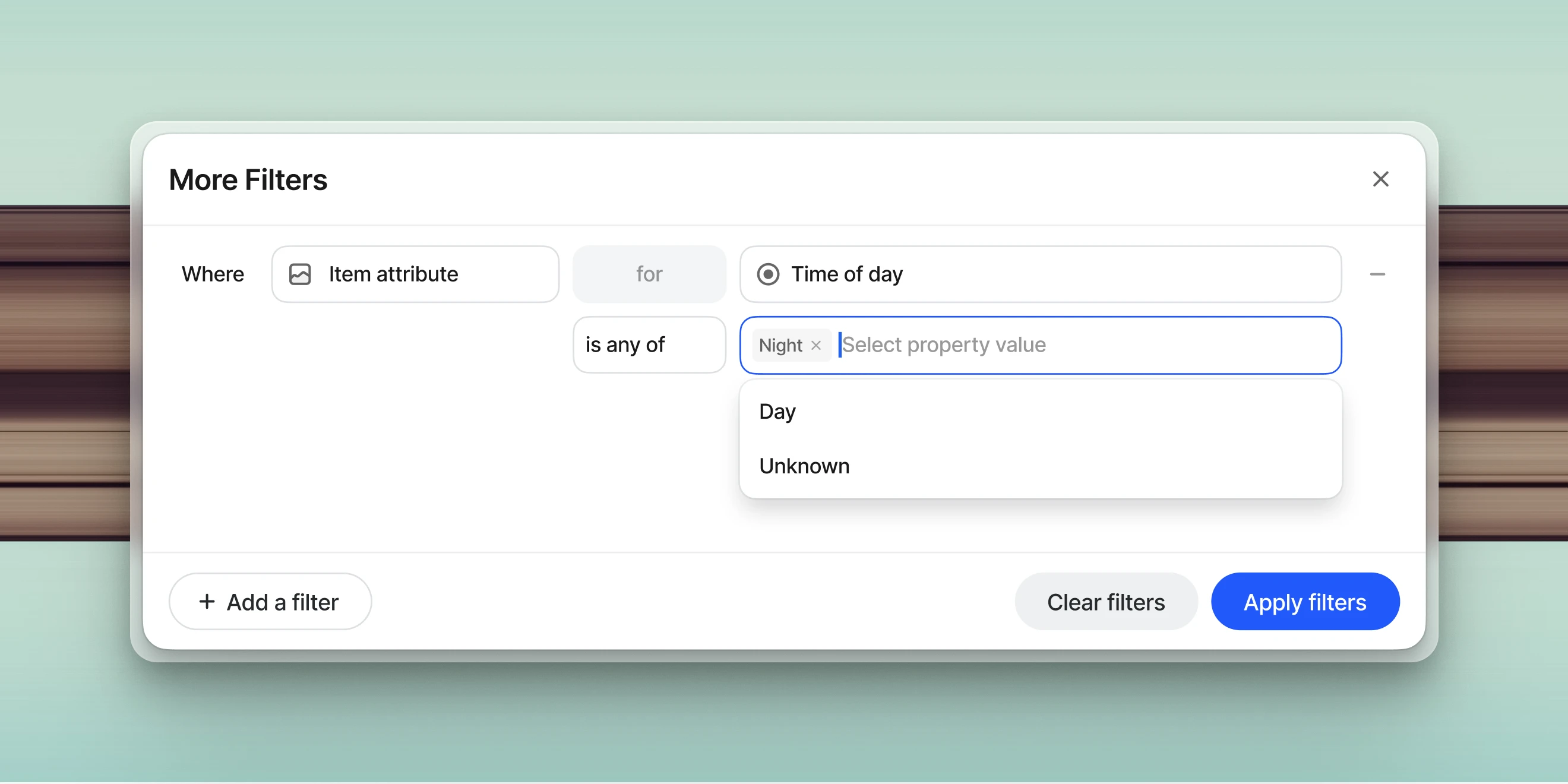
Advanced Filters Now Support Text Properties
Dataset filtering options available in V7 Darwin have just been upgraded. You can now search the full content of text properties to identify whether specific words or phrases have been mentioned. For example, it is possible to automatically generate descriptions of images with AI models and then use them for sorting and filtering files.
You can combine text search with other dataset filters
The search engine for filtering is case-insensitive and will find all instances of a word
To search for multiple words or phrases, add them as multiple filters and set up and/or conditions (you can combine up to 20 filters)
The searched text string should be between 3 and 1000 characters.
This functionality is especially useful for navigating large datasets and pinpointing specific items.
Feb 10, 2025
Item Properties in Logic Stage
You can now use item-level properties as conditions inside any Logic Stage in V7 Darwin. This feature introduces new conditions that let you set up parallel branches in your annotation workflows. For example, if a property is set to a specific value or is missing, an item can be routed back to a previous annotation stage or moved to a custom review stage.
New Conditions:
Verify whether an item property is defined (Item Property is Set/Is Not Set)
Evaluate if an item property contains specific values (Item Property Is Any Of/Is None Of)
To use this functionality, configure the desired properties in your class management tab and then open the workflow editor to add or modify Logic Stages.
Feb 6, 2025
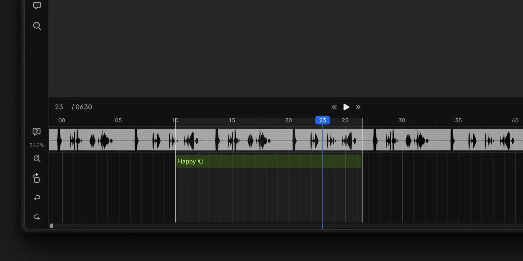
Audiowave Visualization
V7 Darwin now supports Audiowave Visualization. This new feature allows you to overlay an audiowave onto the video timeline. This feature is ideal for use cases such as conversation analysis. You can visualize audio patterns while labeling conversations, which makes the process more intuitive and accurate.
Toggle an audiowave overlay on the video timeline with Shift + A
Use Ctrl + mouse up/down to amplify audio segments for improved clarity
Navigate with smooth playhead movements instead of frame-by-frame jumps
This feature currently supports audio extracted from videos and does not work with audio files. Labels remain tied to video frames rather than specific audio segments, ensuring alignment with existing video-based workflows.
Feb 5, 2025

Oblique Views for Medical Imaging
Oblique Views (also called Rotating Crosshairs) is a new visualization tool for medical imaging professionals working with DICOM files. It enables exploration of 3D structures in Multi-Planar Reconstruction (MPR) DICOMs on non-standard planes.
Key Features
Interactive Crosshairs: Rotate planes with grabbers (dots on crosshairs) using distinct colored lines (red, green, and blue) for each axis.
Oblique Plane Visualization: View MPR DICOMs at non-standard angles.
View Synchronization: Adjusting one plane automatically aligns all views to the same voxel.
3D Mask Support: Visualize 3D masks in oblique views.
How to Use
To use Oblique Views, open a DICOM file in V7 Darwin. Activate the Crosshairs tool using the hotkey R or select it from the interface. On the first use, a brief introductory modal will appear, which you can dismiss permanently. Once active, the crosshairs, with distinct red, green, and blue lines representing each axis, allow you to rotate planes. Adjusting one plane automatically aligns all views to the same voxel for synchronized exploration.
Limitations
Oblique Views is a visualization-only tool and does not support annotation editing. Only 3D masks can be visualized in oblique views, as vertice-based annotations are not supported. Additionally, selecting certain annotation tools, such as the Brush, will revert views to standard planes. This feature is designed to enhance exploration and analysis without altering existing annotations.
Jan 20, 2025
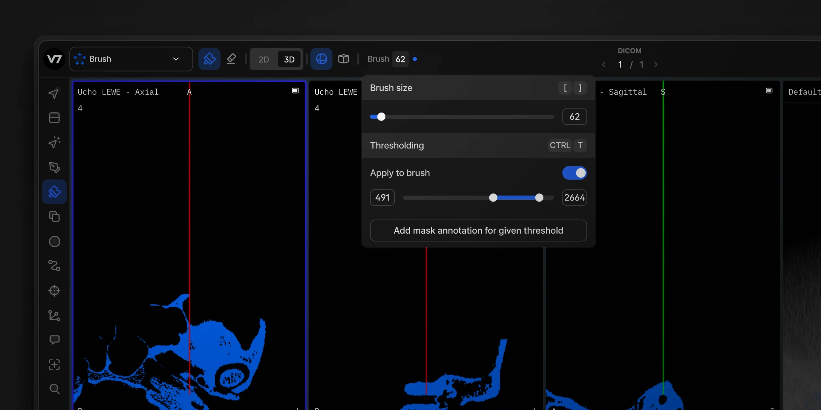
3D Thresholding Brush UI Updates
The 3D thresholding brush now has an improved UI. You can quickly adjust threshold levels and preview which areas of your medical images will be affected by the brush or eraser. Once set, you can annotate with the 3D brush faster than ever and instantly visualize the annotated regions of interest.
Dec 10, 2024
Item-level Attribute Filters in V7 Darwin
Dataset management has just gotten easier with advanced filters for item attributes. Filter your V7 Darwin datasets with multi-select and single-select item attributes. You can add additional criteria and use logical operators to refine the results, which will update instantly.
This update comes in direct response to user feedback, particularly from teams working extensively with item attributes. Support for text-based attributes and filtering with operators like "starts with" and "contains" is currently in development. Stay tuned for more updates coming soon.
Dec 6, 2024